
Chaperone‑Mediated Autophagy in Senescence and Aging
Cellular senescence is a stable cell‑cycle arrest that drives aging and age‑related diseases partly through the senescence‑associated secretory phenotype (SASP). Chaperone‑mediated autophagy (CMA) is a selective lysosomal degradation pathway that maintains proteostasis and influences metabolism, secretion, and immune function. Recent work demonstrates that CMA activity declines with age and that this decline shapes the senescent phenotype, the composition of the SASP, and the ability of the immune system to clear senescent cells.
Introduction
Aging is accompanied by a progressive loss of proteostasis, a hallmark of which is the downregulation of CMA. CMA degrades proteins containing a KFERQ‑like motif via the lysosomal receptor LAMP2A. In young cells, CMA is upregulated during the onset of senescence, but this response is blunted in cells from old organisms. Understanding how CMA deficiency alters senescence and its downstream effects is essential for developing interventions that target age‑related decline.
CMA Decline in Aged Cells and Senescence Induction
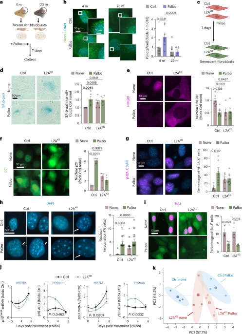
Using primary mouse ear fibroblasts (mEFs) from young (4 mo) and aged (23 mo) mice, researchers found that aged fibroblasts exhibit lower basal CMA activity and fail to upregulate CMA after induction of senescence with palbociclib. This impairment is not due to reduced expression of the CMA reporter but reflects defective lysosomal delivery. Knockdown of Lamp2a (L2AKD) in young NIH3T3 fibroblasts recapitulates several senescence markers (SA‑β‑gal, nuclear HMGB1 loss, p21 upregulation) without inducing full proliferative arrest, indicating that CMA loss alone does not drive conventional senescence.
Proteomic and Metabolic Consequences of CMA Loss
Comparative proteomics of young control, young CMA‑deficient (L2AKO), and aged control mEFs revealed that CMA loss reproduces ~45 % of the basal proteome changes seen in aged cells. Pathway analysis enriched for lipid metabolism, oxidative phosphorylation, translation, vesicular trafficking, and ECM remodeling. Upon senescence induction, proteins that normally decrease—involved in translation, immune signaling, glucose metabolism, and actin dynamics—fail to decline in CMA‑deficient cells.
Pathway Enrichment
| Pathway | Change in Aged/CMA‑KO |
|---|---|
| Lipid metabolism | ↑ Altered enzyme expression |
| Oxidative phosphorylation | ↓ Reduced complex activity |
| Vesicular trafficking | ↑ Accumulation of trafficking proteins |
| ECM remodeling | ↑ Increased matrix proteins |
Metabolomic profiling showed that CMA‑deficient cells mimic many basal metabolic features of aged cells, including altered energy production, carbohydrate and lipid metabolism, and amino‑acid pathways. During senescence, young control cells engage fatty acid β‑oxidation, whereas CMA‑deficient and aged cells fail to do so, instead activating the TCA cycle and nucleotide metabolism—a pattern linked to defective CMA‑dependent degradation of metabolic enzymes.
Secretome Alterations and Paracrine Senescence
The SASP is composed of conventionally and unconventionally secreted proteins. Using ER‑TurboID biotinylation, the study separated these fractions. CMA‑knockdown cells under basal conditions showed increased secretion of ECM, migration, and antigen‑presentation proteins, and decreased secretion of inflammation and lipid‑metabolism factors. Unconventional secretion of nuclear proteins (e.g., chromosome organization, DNA repair) was paradoxically elevated.
Paracrine Effects
The secretome of CMA‑deficient fibroblasts induces paracrine senescence in neighboring cells, evidenced by increased SA‑β‑gal activity, p21 nuclear localization, and loss of nuclear HMGB1.
When senescence was induced, CMA‑deficient cells failed to downregulate many conventionally secreted proteins and instead upregulated factors involved in cell migration, ER folding, and immune activation. The resulting secretome had a stronger pro‑inflammatory profile and enhanced the ability to spread senescence to adjacent cells.
Impact on Macrophage Function and Immune Clearance
Bone‑marrow‑derived macrophages (BMDMs) from aged mice exhibit reduced CMA activity. Exposure to the secretome of senescent fibroblasts further suppresses macrophage CMA, suggesting a feed‑forward loop that impairs immune surveillance. In co‑culture experiments, macrophages lacking LAMP2A (M‑L2A−/−) showed decreased clearance of senescent fibroblasts and increased cytotoxicity when exposed to CMA‑deficient senescent cells.
Phagocytosis Defect
CMA‑deficient macrophages display reduced phagocytic and efferocytic capacities. This defect is linked to impaired lysosomal degradation of the “don’t‑eat‑me” signal SIRPα, leading to its accumulation and sustained activation of SHP‑1, which inhibits actin cytoskeleton reorganization required for phagocytosis. The secretome from CMA‑deficient senescent cells further diminishes macrophage phagocytosis, an effect partially rescued by the CMA activator CA77.1.
Therapeutic Activation of CMA Reduces Senescence Burden
Oral administration of the small‑molecule CMA activator CA77.1 (30 mg kg⁻¹) to 18‑month‑old mice for five months prevented the age‑associated rise in SA‑β‑gal⁺ cells in gonadal white adipose tissue, liver, and lung, and reduced expression of senescence markers p21, Tnf, Il1α, and p16INK4A. Histological analysis showed decreased fibrosis in liver and adipose tissue. Principal component analysis confirmed that CA‑treated aged mice clustered closer to young mice.
In vitro, CA77.1 restored the phagocytic activity of BMDMs from aged mice to levels comparable to those from young mice, indicating that part of the benefit is mediated by improved macrophage function.
CMA Activation in Lung Fibrosis
Analysis of human lung single‑cell RNA‑seq data revealed a decline in a transcriptional CMA score with age in fibroblasts, alveolar type 1 and type 2 cells, and specific macrophage subsets. Patients with idiopathic pulmonary fibrosis (IPF) exhibited lower CMA scores and reduced LAMP2A protein stability in lysosomes. In a mouse model of bleomycin‑induced fibrosis, early administration of CA77.1 (starting day 2) preserved body weight, reduced collagen deposition, and lowered expression of fibrosis and senescence genes. Immune profiling showed that early CMA activation normalized macrophage numbers and promoted a resolving immune environment, whereas later treatment was less effective.
Conclusion
The cumulative evidence positions CMA as a critical regulator of senescence, SASP composition, and immune clearance. Age‑related CMA decline drives a senescence‑like phenotype, alters the secretome, and impairs macrophage‑mediated removal of senescent cells. Pharmacological activation of CMA with CA77.1 mitigates these defects, reducing senescence burden and fibrosis in multiple organs. These findings highlight CMA as a promising therapeutic target for age‑related diseases, including idiopathic pulmonary fibrosis.
Science, Health, Aging, Cellular Senescence, Chaperone‑Mediated Autophagy, Macrophages, Immunology, Pharmacology

